
Page 150 - Demo
P. 150
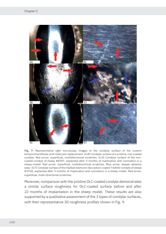
Chapter 5148Fig. 7: Representative light microscopy images of the condylar surface of the custom temporomandibular joint total joint replacement. (A,B) Condylar surface of a pristine, non-coated condyle. Red arro superficial, multidirectional scratches. (C,D) Condylar surface of the noncoated condyle of sheep #8787, explanted after 9 months of mastication and rumination in a sheep model. Red arro superficial, multidirectional scratches. Blue arro deeper abrasive wear. (E,F) Condylar surface of the HadSat diamond-like carbon-coated Ti6Al4V condyle of sheep #5158, explanted after 9 months of mastication and rumination in a sheep model. Red arro superficial, multi-directional scratches.Moreover, comparison with the pristine DLC-coated condyle demonstrates a similar surface roughness for DLC-coated surface before and after 22 months of implantation in the sheep model. These results are also supported by a qualitative assessment of the 3 types of condylar surfaces, with their representative 3D roughness profiles shown in Fig. 9. Nikolas de Meurechy NW.indd 148 05-06-2024 10:14
