
Page 145 - Demo
P. 145
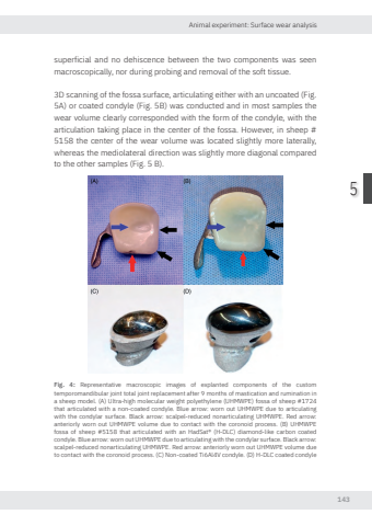
Animal experiment: Surface wear analysis1435superficial and no dehiscence between the two components was seen macroscopically, nor during probing and removal of the soft tissue. 3D scanning of the fossa surface, articulating either with an uncoated (Fig. 5A) or coated condyle (Fig. 5B) was conducted and in most samples the wear volume clearly corresponded with the form of the condyle, with the articulation taking place in the center of the fossa. However, in sheep # 5158 the center of the wear volume was located slightly more laterally, whereas the mediolateral direction was slightly more diagonal compared to the other samples (Fig. 5 B). Fig. 4: Representative macroscopic images of explanted components of the custom temporomandibular joint total joint replacement after 9 months of mastication and rumination in a sheep model. (A) Ultra-high molecular weight polyethylene (UHMWPE) fossa of sheep #1724 that articulated with a non-coated condyle. Blue arro worn out UHMWPE due to articulating with the condylar surface. Black arro scalpel-reduced nonarticulating UHMWPE. Red arro anteriorly worn out UHMWPE volume due to contact with the coronoid process. (B) UHMWPE fossa of sheep #5158 that articulated with an HadSat® (H-DLC) diamond-like carbon coated condyle. Blue arro worn out UHMWPE due to articulating with the condylar surface. Black arro scalpel-reduced nonarticulating UHMWPE. Red arro anteriorly worn out UHMWPE volume due to contact with the coronoid process. (C) Non-coated Ti6Al4V condyle. (D) H-DLC coated condyleNikolas de Meurechy NW.indd 143 05-06-2024 10:14
