
Page 139 - Demo
P. 139
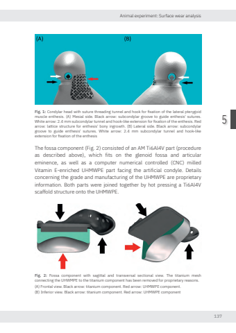
Animal experiment: Surface wear analysis1375Fig. 1: Condylar head with suture threading tunnel and hook for fixation of the lateral pterygoid muscle enthesis. (A) Mesial side. Black arro subcondylar groove to guide enthesis' sutures. White arro 2.4 mm subcondylar tunnel and hook-like extension for fixation of the enthesis. Red arro lattice structure for enthesis' bony ingrowth. (B) Lateral side. Black arro subcondylar groove to guide enthesis' sutures. White arro 2.4 mm subcondylar tunnel and hook-like extension for fixation of the enthesisThe fossa component (Fig. 2) consisted of an AM Ti6Al4V part (procedure as described above), which fits on the glenoid fossa and articular eminence, as well as a computer numerical controlled (CNC) milled Vitamin E-enriched UHMWPE part facing the artificial condyle. Details concerning the grade and manufacturing of the UHMWPE are proprietary information. Both parts were joined together by hot pressing a Ti6Al4V scaffold structure onto the UHMWPE. Fig. 2: Fossa component with sagittal and transversal sectional view. The titanium mesh connecting the UHWMPE to the titanium component has been removed for proprietary reasons. (A) Frontal view. Black arro titanium component. Red arro UHMWPE component.(B) Inferior view. Black arro titanium component. Red arro UHMWPE componentNikolas de Meurechy NW.indd 137 05-06-2024 10:14
