
Page 126 - Demo
P. 126
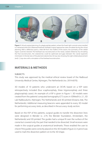
124Chapter7Figure1VirtualsurgicalplanningofaplagiocephalypatientwherethefusedrightcoronalsutureresultedinaretractedorbitandaflattenedforeheadMultiplesurgicalapproachesweresimulatedduringthevirtualplanningWithadeflectionpointalittleleftfromthemidlineofthenasionthesymmetryoftheorbitalregioncouldberestoredTheforeheadwasreconstructedonthenewlycreatedorbitalbarBysimulatingthefinalreconstructiontheosteotomiescouldbeoptimizedandgapsbetweenthecranialsegmentsminimizedA)frontalviewoftheplannedosteotomiesB)topviewshowingtherightorbitaladvancement(red)C)topviewwithasimulationoftheforeheadreconstructionMATERIALS&METHODSSUBJECTSThisstudywasapprovedbythemedicalethicalreviewboardoftheRadboudUniversityMedicalCentreNijmegen TheNetherlands(no2019-6070)3Dmodelsof10patientswhounderwentanOCVRbasedonaVSPwereretrospectivelyincluded(fourscaphocephalythreetrigonocephalyandthreeplagiocephalycases)AnexampleofaVSPisgiveninFigure13Dmodelswerecreated from thepatients’computed tomography(CT)scansin3DMedX(v213DLabRadboudumcNijmegen TheNetherlands)and3Dprinted(OceanzEde TheNetherlands)Additionalmeasuringbeaconswereappendedtoevery3DmodelforperformingaccuracytestsasdescribedintheaccuracystudysectionBasedontheVSPofthepatientssurgicalguidestotransferthedissectionlinesweredesignedinBlender(v279theBlenderfoundationAmsterdamtheNetherlands)and3Dprinted TheguideshadauniquefitoverthesurfaceofthecraniabutcoveredonlythepartthatneededtobedissectedSmallrecessesweremadeinthesurgicalguidesatanatomicallandmarksTheseenabledtheusertocheckif theguides werecorrectlyplacedon the3Dmodels(Figure2) Apencil wasusedtomarkthedissectionpatternontothe3Dshape
