
Page 16 - Advanced concepts in orbital wall fractures
P. 16
14
Chapter 1
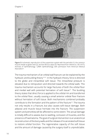
Figure 3 A schematic reproduction of the suspension system with the periorbit (1), the common muscle sheath at globe level (2), and the fibrous septa (3). Reprinted from reference 2 © (1977), Archives of Ophthalmology / JAMA Ophthalmology, with permission from American Medical Association2.
The trauma mechanism of an orbital wall fracture can be explained by the hydraulic and buckling theory13,14. In the hydraulic theory, force is directed to the globe and intraorbital soft tissue. The intraorbital pressure is elevated due to retropulsion and directed towards the orbital walls. This trauma mechanism accounts for large fractures of both the orbital floor and medial wall with potential herniation of soft tissue15. The buckling theory states that direct force is applied to the orbital rim and transferred to the orbital floor, usually causing a small anterior orbital floor fracture without herniation of soft tissue. Both mechanisms combined probably contribute to the formation and the pattern of the fracture14. The trauma not only results in a fracture, but also causes soft tissue damage. Both adipose and muscle tissue herniate into the fracture. The suspension system and periorbita will be affected to some extent. The actual damage is initially difficult to assess due to swelling, contusion of muscles, and the presence of haematoma. The goal of surgical intervention is an anatomical reconstruction of the bony walls and the release of incarcerated soft tissue to restore orbital function. The regenerative capacity of the soft tissue and the amount of damage caused by the surgery itself is unpredictable.
