
Page 14 - Advanced concepts in orbital wall fractures
P. 14
Chapter 1
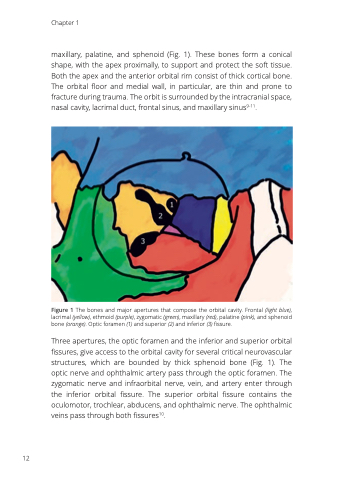
maxillary, palatine, and sphenoid (Fig. 1). These bones form a conical shape, with the apex proximally, to support and protect the soft tissue. Both the apex and the anterior orbital rim consist of thick cortical bone. The orbital floor and medial wall, in particular, are thin and prone to fracture during trauma. The orbit is surrounded by the intracranial space, nasal cavity, lacrimal duct, frontal sinus, and maxillary sinus9-11.
Figure 1 The bones and major apertures that compose the orbital cavity. Frontal (light blue), lacrimal (yellow), ethmoid (purple), zygomatic (green), maxillary (red), palatine (pink), and sphenoid bone (orange). Optic foramen (1) and superior (2) and inferior (3) fissure.
Three apertures, the optic foramen and the inferior and superior orbital fissures, give access to the orbital cavity for several critical neurovascular structures, which are bounded by thick sphenoid bone (Fig. 1). The optic nerve and ophthalmic artery pass through the optic foramen. The zygomatic nerve and infraorbital nerve, vein, and artery enter through the inferior orbital fissure. The superior orbital fissure contains the oculomotor, trochlear, abducens, and ophthalmic nerve. The ophthalmic veins pass through both fissures10.
12
