
Page 15 - Advanced concepts in orbital wall fractures
P. 15
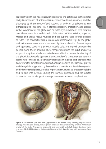
                                Together with these neurovascular structures, the soft tissue in the orbital C1 cavity is composed of adipose tissue, connective tissue, muscles, and the
globe (Fig. 2). The majority of soft tissue is fat and can be subdivided into
extraconal and intraconal fat. It provides support, protection, and assists
in the movement of the globe and muscles. Eye movement, with rotation over three axes, is a well-timed collaboration of the inferior, superior, medial, and lateral rectus muscles and the superior and inferior oblique muscles. The connective tissue is a complex framework (Fig. 3). The globe and extraocular muscles are enclosed by fascia sheaths. Several septa and ligaments, containing smooth muscle cells, are aligned between the periorbit and these sheaths. They compartmentalize the orbit and are a suspension system which seems to be crucial to the normal functioning of the globe2. Lockwood’s ligament is an example of a transverse suspensory ligament for the globe. It vertically stabilizes the globe and provides the framework for the inferior rectus and oblique muscles. The lacrimal system and the eyelids, supported by the medial and lateral canthi and the superior and inferior tarsal plates, are also important structures to protect the globe and to take into account during the surgical approach and the orbital reconstruction, as iatrogenic damage can cause serious complications.
Figure 2 The coronal (left) and axial (right) view of the orbital cavity showing adipose tissue (orange), muscles (red striped), nerves (yellow and white tubular structures), arteries (red tubular structures), the globe (transparent and white), and bony orbit and skull (beige). Reprinted from reference 12 © (2019), Atlas of Endoscopic Sinus and Skull Base Surgery, with permission from Elsevier12.
General introduction and outline
  13
