
Page 41 - Effects of radiotherapy and hyperbaric oxygen therapy on oral microcirculation Renee Helmers
P. 41
Hyperoxia-driven microvascular changes
Interestingly, there were no statistically significant differences observed for Øbv
at any HB time points or return to baseline conditions. MicroScan recordings
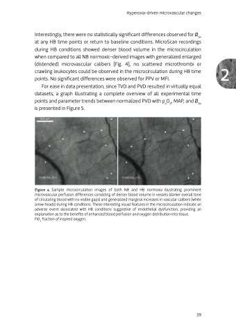
during HB conditions showed denser blood volume in the microcirculation
when compared to all NB normoxic-derived images with generalized enlarged
(distended) microvascular calibers [Fig. 4], no scattered microthrombi or 2 crawling leukocytes could be observed in the microcirculation during HB time
points. No significant differences were observed for PPV or MFI.
For ease in data presentation, since TVD and PVD resulted in virtually equal datasets, a graph illustrating a complete overview of all experimental time points and parameter trends between normalized PVD with paO2, MAP, and Øbv
is presented in Figure 5.
Figure 4. Sample microcirculation images of both NB and HB normoxia illustrating prominent microvascular perfusion differences consisting of denser blood volume in vessels (darker overall tone of circulating blood with no visible gaps) and generalized marginal increases in vascular calibers (white arrow heads) during HB conditions. These interesting visual features in the microcirculation indicate an adverse event associated with HB conditions suggestive of endothelial dysfunction, providing an explanation as to the benefits of enhanced blood perfusion and oxygen distribution into tissue.
FiO2 fraction of inspired oxygen.
39
