
Page 70 - Bladder Dysfunction in the Context of the Bladder-Brain Connection - Ilse Groenendijk.pdf
P. 70
68
Chapter 3
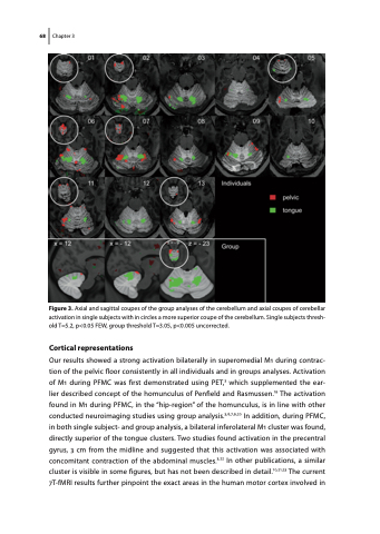
Figure 3. Axial and sagittal coupes of the group analyses of the cerebellum and axial coupes of cerebellar activation in single subjects with in circles a more superior coupe of the cerebellum. Single subjects thresh- old T=5.2, p<0.05 FEW, group threshold T=3.05, p<0.005 uncorrected.
Cortical representations
Our results showed a strong activation bilaterally in superomedial M1 during contrac- tion of the pelvic floor consistently in all individuals and in groups analyses. Activation of M1 during PFMC was first demonstrated using PET,3 which supplemented the ear- lier described concept of the homunculus of Penfield and Rasmussen.19 The activation found in M1 during PFMC, in the “hip-region” of the homunculus, is in line with other conducted neuroimaging studies using group analysis.3,6,7,9,20 In addition, during PFMC, in both single subject- and group analysis, a bilateral inferolateral M1 cluster was found, directly superior of the tongue clusters. Two studies found activation in the precentral gyrus, 3 cm from the midline and suggested that this activation was associated with concomitant contraction of the abdominal muscles.3,22 In other publications, a similar cluster is visible in some figures, but has not been described in detail.10,21,23 The current 7T-fMRI results further pinpoint the exact areas in the human motor cortex involved in
