
Page 67 - Bladder Dysfunction in the Context of the Bladder-Brain Connection - Ilse Groenendijk.pdf
P. 67
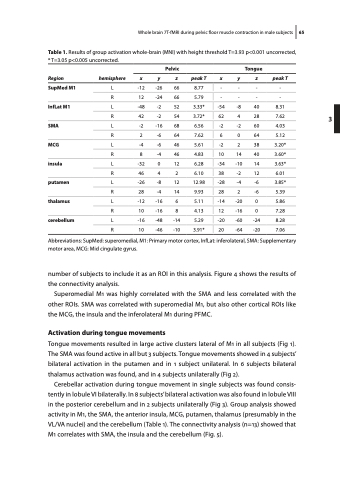
                 Whole brain 7T-fMRI during pelvic floor muscle contraction in male subjects 65 Table 1. Results of group activation whole-brain (MNI) with height threshold T=3.93 p<0.001 uncorrected,
* T=3.05 p<0.005 uncorrected.
 Region
 hemisphere
 Pelvic Tongue
 x
 y
 z
 peak T x
 y
 z
 peak T
 SupMed M1
 L
 -12
 -26
 66
  8.77 -
 -
 -
 -
 R
  12
  -24
  66
    5.79 -
    -
  -
  -
  InfLat M1
L
-48
-2
52
3.33* -54
-8
40
8.31
 R
 42
 -2
 54
   3.72* 62
 4
 28
 7.62
 SMA
 L
  -2
  -16
  68
    6.56 -2
    -2
  60
  4.03
 R
2
-6
64
7.62 6
0
64
5.12
 MCG
  L
  -4
  -6
  46
    5.61 -2
    2
  38
  3.20*
 R
8
-4
46
4.83 10
14
40
3.60*
insula
 L
 -32
 0
 12
   6.28 -34
 -10
 14
 3.63*
 R
 46
 4
 2
   6.10 38
 -2
 12
 6.01
 putamen
  L
  -26
  -8
  12
    12.98 -28
    -4
  -6
  3.85*
 R
28
-4
14
9.93 28
2
-6
5.39
thalamus
  L
 -12
 -16
 6
   5.11 -14
 -20
 0
 5.86
 R
 10
 -16
 8
   4.13 12
 -16
 0
 7.28
cerebellum
  L
  -16
  -48
  -14
    5.29 -20
    -60
  -24
  8.28
 R
 10
 -46
 -10
 3.91* 20
  -64
 -20
 7.06
     Abbreviations: SupMed: superomedial, M1: Primary motor cortex, InfLat: inferolateral, SMA: Supplementary motor area, MCG: Mid cingulate gyrus.
number of subjects to include it as an ROI in this analysis. Figure 4 shows the results of the connectivity analysis.
Superomedial M1 was highly correlated with the SMA and less correlated with the other ROIs. SMA was correlated with superomedial M1, but also other cortical ROIs like the MCG, the insula and the inferolateral M1 during PFMC.
Activation during tongue movements
Tongue movements resulted in large active clusters lateral of M1 in all subjects (Fig 1). The SMA was found active in all but 3 subjects. Tongue movements showed in 4 subjects’ bilateral activation in the putamen and in 1 subject unilateral. In 6 subjects bilateral thalamus activation was found, and in 4 subjects unilaterally (Fig 2).
Cerebellar activation during tongue movement in single subjects was found consis- tently in lobule VI bilaterally. In 8 subjects’ bilateral activation was also found in lobule VIII in the posterior cerebellum and in 2 subjects unilaterally (Fig 3). Group analysis showed activity in M1, the SMA, the anterior insula, MCG, putamen, thalamus (presumably in the VL/VA nuclei) and the cerebellum (Table 1). The connectivity analysis (n=13) showed that M1 correlates with SMA, the insula and the cerebellum (Fig. 5).
3
