
Page 159 - Quantitative Imaging of Small Tumours with Positron Emission Tomography
P. 159
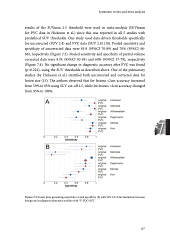
Chapter 7 Figure 7.4: Summary ROC curves of discrimination between benign and malignant pulmonary nodules with 18F-FDG-PET. With diagnosis of breast lesions, using data-driven SUVmean thresholds of 2.1 for PVC and non-PVC, at a fixed specificity of 90%, PVC increased sensitivity from 69 to 81%, but the impact on accuracy was not statistically significant (21). In discriminating between aggressive and indolent non-Hodgkin lymphoma (NHL), PVC decreased specificity without affecting sensitivity (22). Similarly, PVC did not improve differentiating between high- and low-grade NHL (23). PVC to enabled differentiation between indolent NHL and Kikuchi-Fujimoto disease (24). Staging Studies evaluating the effect of PVC on staging (Table 7.2, n=10) included lung (n=3), breast (n=2), thyroid (n=1), head-and-neck squamous cell (n=1), nasopharyngeal (n=1), prostate (n=1), and colorectal cancer (n=1). Applied PVC methods were the recovery coefficient-method (n=4), PSF reconstruction (n=4), iterative deconvolution (n=1) and geometric transfer matrix (n=1). Most of these studies did not specify SUV thresholds of test positivity for uncorrected and PVC data. Four studies did not specify lesions sizes. One study stratified both uncorrected and PVC data for lesion size in secondary analysis. In non-small cell lung cancer (NSCLC) patients the association between primary tumour SUVmax and overall TNM stage disappeared after PVC (25). 158
