
Page 111 - Helicobacter pylori and Gastric Cancer: From Tumor microenvironment to Immunotherapy
P. 111
Endoscopy examination
Helicobacter pylori
Helicobacter Pylori
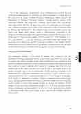
All of the endoscopic examinations were performed and assessed by one experienced endoscopist (G.A and K.D) who had been trained to evaluate EGA at the University of Abuja Teaching Hospital, Gwagwalada. Abuja. Nigeria and Department of Medical Laboratory Sciences. Marand Branch, Islamic Azad University, Marand, Iran respectively. Olympus video-scopes with conventional white light (model GIF-160; Olympus) were used. The endoscopic mucosal atrophy was evaluated according to the location of the endoscopic atrophic border described by Kimura and Takemoto[17]. This atrophic border is the boundary between the pyloric and fundic gland regions, which is endoscopically recognized by the difference in color and height of the gastric mucosa between the two sides of the border (Fig. 1). There are three grades of EGA: severe (O2 – O3), moderate (C3 – O1), and mild (C1 – C2). Six specimens were taken from each patient: five specimens were taken from specific locations according to the updated Sydney System and were put in separate boxes for pathologic examination; the 6th specimen used for rapid urease test was taken from the greater curvature of the antrum.
Level of agreement definition
The endoscopic findings of the extent of atrophy were compared with the histological findings of glandular atrophy at five biopsy sites (Figure 1). To be able to compare the extent of atrophy strictly, both classifications were modified to five grades according to definitions of those anatomical locations. Histological grading was scored as 1, none; 2, antrum (site 1 and/or 2); 3, angulus (up to site 3); 4, the middle body of the lesser curvature (up to site 4) and 5, the middle body of the greater curvature (up to site 5). Endoscopic atrophic grading according to the modified Kimura–Takemoto classification was scored as 1, none; 2; antral (C-1); 3, antral predominant (C-2); 4, corpus predominant (C-3, O-1, O-2) and 5, pan-atrophy (O-3) (Figure 3). Inasmuch as extensive atrophy is associated with a much higher cancer risk than limited atrophy, the Kimura–Takemoto classification was simplified to three grades of cancer risk oriented atrophy as: normal (no atrophy), limited atrophy (antral and antral predominant atrophy; C-1, C-2) and extended atrophy (corpus predominant and pan-atrophy; C-3, O-1, O-2, O-3). Agreement was defined as matching of endoscopic and histologic grades, with all other findings defined as disagreement.
105
109
5
