
Page 113 - Helicobacter pylori and Gastric Cancer: From Tumor microenvironment to Immunotherapy
P. 113
Helicobacter Pylori
Helicobacter pylori
RESULTS
Demographic data
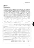
A total of 248 patients aged 50±30 years were included in this study, including 124 patients from Nigeria and 124 from Iran. Mean ± SD patient age was 46.4 ( ± 15.3) years. Of these patients, 131(52.8%) were male, 117 female;(47.2%). Gender ratio F/M :0.89 and 138 (55.6%) were serologically H. pylori-positive. The detailed characteristics of the two subgroups are shown in Table 1. There were significant differences between the groups from the Nigeria and Iran, especially with respect to the extent of atrophy. In the Nigerian population, only 60.% were diagnosed histologically of having corpus atrophy, whereas, in Iran, 54% had gastric atrophy while 57 (46.0%) showed no evidence of histological atrophy. Hence geographical origin influences disease presentation necessitating comparison of the relative performance of endoscopy with respect to the diagnosis of AG.
Table 1: Demographic, endoscopic, and pathologic characteristics of study population with H pylori associated gastric atrophy.
Nigeria (n = 124) (%)
Iran (n =124) (%)
Total (n =248) (%)
P value (Nigeria vs Iran)
Clinical parameters
Sex
Male
62 (50.0)
69 (55.6)
131 (52.8)
0.37
Female
62 (50.0)
55 (44.4)
117 (47.2)
Age (yr)
≥ 40
69 (55.6)
68 (54.8)
137 (55.2)
0.89
< 40
55 (44.4)
56 (45.2)
111 (44.8)
Helicobacter pylori Ag
Positive
40 (32.3)
88 (71.0)
128 (51.6)
0.84
Negative
84 (67.7)
36 (29.0)
120 (48.0)
Helicobacter pylori IgG
Positive
89 (71.8)
49 (39.5)
138 (55.6)
0.00
Negative
35 (28.2)
75 (60.5)
112 (45.4)
Serologic features (ng/mL), mean ± SD
Pepsinogen I
49.9 ± 39.2
57.8 ± 38.1
53.1 ± 38.7
0.731
Pepsinogen II
12.9 ± 7.2
12.1 ± 10.1
12.9 ± 8.65
0.621
107
111
5
