
Page 26 - The role of advanced echocardiography in patients with ischemic heart disease - Rachid Abou
P. 26
Chapter one. General introduction and outline of the thesis
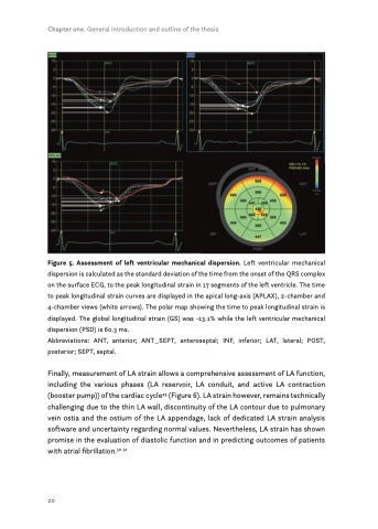
Figure 5. Assessment of left ventricular mechanical dispersion. Left ventricular mechanical dispersion is calculated as the standard deviation of the time from the onset of the QRS complex on the surface ECG, to the peak longitudinal strain in 17 segments of the left ventricle. The time to peak longitudinal strain curves are displayed in the apical long-axis (APLAX), 2-chamber and 4-chamber views (white arrows). The polar map showing the time to peak longitudinal strain is displayed. The global longitudinal strain (GS) was -13.1% while the left ventricular mechanical dispersion (PSD) is 60.3 ms.
Abbreviations: ANT, anterior; ANT_SEPT, anteroseptal; INF, inferior; LAT, lateral; POST, posterior; SEPT, septal.
Finally, measurement of LA strain allows a comprehensive assessment of LA function, including the various phases (LA reservoir, LA conduit, and active LA contraction (booster pump)) of the cardiac cycle49 (Figure 6). LA strain however, remains technically challenging due to the thin LA wall, discontinuity of the LA contour due to pulmonary vein ostia and the ostium of the LA appendage, lack of dedicated LA strain analysis software and uncertainty regarding normal values. Nevertheless, LA strain has shown promise in the evaluation of diastolic function and in predicting outcomes of patients with atrial fibrillation.50-51
20
