
Page 25 - The role of advanced echocardiography in patients with ischemic heart disease - Rachid Abou
P. 25
1
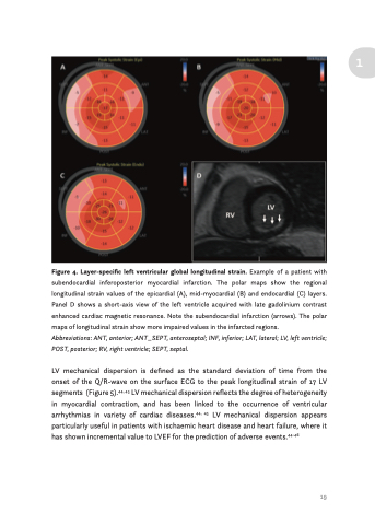
Figure 4. Layer-specific left ventricular global longitudinal strain. Example of a patient with subendocardial inferoposterior myocardial infarction. The polar maps show the regional longitudinal strain values of the epicardial (A), mid-myocardial (B) and endocardial (C) layers. Panel D shows a short-axis view of the left ventricle acquired with late gadolinium contrast enhanced cardiac magnetic resonance. Note the subendocardial infarction (arrows). The polar maps of longitudinal strain show more impaired values in the infarcted regions.
Abbreviations: ANT, anterior; ANT_SEPT, anteroseptal; INF, inferior; LAT, lateral; LV, left ventricle; POST, posterior; RV, right ventricle; SEPT, septal.
LV mechanical dispersion is defined as the standard deviation of time from the onset of the Q/R-wave on the surface ECG to the peak longitudinal strain of 17 LV segments (Figure 5).44, 45 LV mechanical dispersion reflects the degree of heterogeneity in myocardial contraction, and has been linked to the occurrence of ventricular arrhythmias in variety of cardiac diseases.44, 45 LV mechanical dispersion appears particularly useful in patients with ischaemic heart disease and heart failure, where it has shown incremental value to LVEF for the prediction of adverse events.44-46
19
