
Page 16 - Demo
P. 16
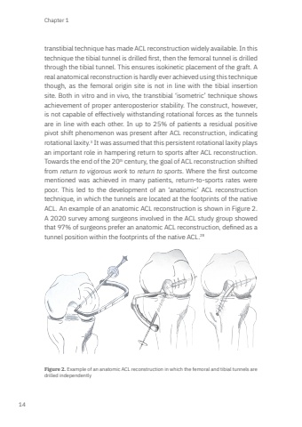
Chapter 114transtibial technique has made ACL reconstruction widely available. In this technique the tibial tunnel is drilled first, then the femoral tunnel is drilled through the tibial tunnel. This ensures isokinetic placement of the graft. A real anatomical reconstruction is hardly ever achieved using this technique though, as the femoral origin site is not in line with the tibial insertion site. Both in vitro and in vivo, the transtibial ‘isometric’ technique shows achievement of proper anteroposterior stability. The construct, however, is not capable of effectively withstanding rotational forces as the tunnels are in line with each other. In up to 25% of patients a residual positive pivot shift phenomenon was present after ACL reconstruction, indicating rotational laxity.3 It was assumed that this persistent rotational laxity plays an important role in hampering return to sports after ACL reconstruction. Towards the end of the 20th century, the goal of ACL reconstruction shifted from return to vigorous work to return to sports. Where the first outcome mentioned was achieved in many patients, return-to-sports rates were poor. This led to the development of an ‘anatomic’ ACL reconstruction technique, in which the tunnels are located at the footprints of the native ACL. An example of an anatomic ACL reconstruction is shown in Figure 2. A 2020 survey among surgeons involved in the ACL study group showed that 97% of surgeons prefer an anatomic ACL reconstruction, defined as a tunnel position within the footprints of the native ACL.28Figure 2. Example of an anatomic ACL reconstruction in which the femoral and tibial tunnels are drilled independentlyMark Zee.indd 14 03-01-2024 08:56
