
Page 138 - Demo
P. 138
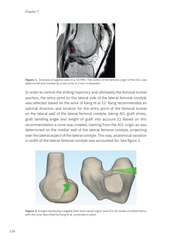
Chapter 7136Figure 1. Example of sagittal view of a 3D MRI. The center of the femoral origin of the ACL was determined and marked by a red circle of 2 mm in diameterIn order to control the drilling trajectory and ultimately the femoral tunnel position, the entry point on the lateral side of the lateral femoral condyle was selected based on the work of Kang et al.12 Kang recommended an optimal direction and location for the entry point of the femoral tunnel on the lateral wall of the lateral femoral condyle, taking ACL graft stress, graft bending angle and length of graft into account.12 Based on this recommendation a cone was created, starting from the ACL origin as was determined on the medial wall of the lateral femoral condyle, projecting over the lateral aspect of the lateral condyle. This way, anatomical variation in width of the lateral femoral condyle was accounted for. See figure 2. Figure 2. Images displaying a sagittal (left) and cranial (right) view of a 3D model of a distal femur with the cone described by Kang et al. projected in placeMark Zee.indd 136 03-01-2024 08:56
