
Page 67 - Development of Functional Scaffolds for Bone Tissue Engineering Using 3D-Bioprinting of Cells and Biomaterials - Yasaman Zamani
P. 67
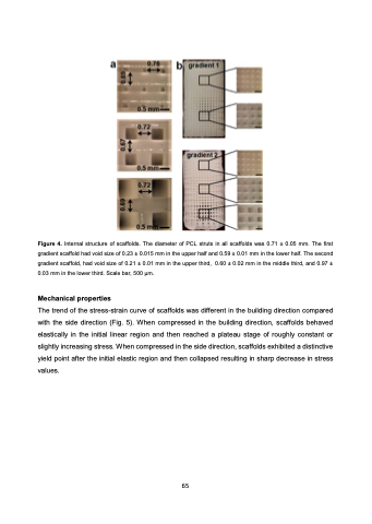
 Figure 4. Internal structure of scaffolds. The diameter of PCL struts in all scaffolds was 0.71 ± 0.05 mm. The first gradient scaffold had void size of 0.23 ± 0.015 mm in the upper half and 0.59 ± 0.01 mm in the lower half. The second gradient scaffold, had void size of 0.21 ± 0.01 mm in the upper third, 0.60 ± 0.02 mm in the middle third, and 0.97 ± 0.03 mm in the lower third. Scale bar, 500 μm.
Mechanical properties
The trend of the stress-strain curve of scaffolds was different in the building direction compared with the side direction (Fig. 5). When compressed in the building direction, scaffolds behaved elastically in the initial linear region and then reached a plateau stage of roughly constant or slightly increasing stress. When compressed in the side direction, scaffolds exhibited a distinctive yield point after the initial elastic region and then collapsed resulting in sharp decrease in stress values.
65
