
Page 115 - Development of Functional Scaffolds for Bone Tissue Engineering Using 3D-Bioprinting of Cells and Biomaterials - Yasaman Zamani
P. 115
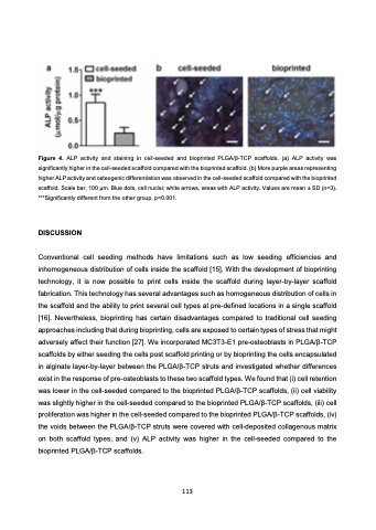
 Figure 4. ALP activity and staining in cell-seeded and bioprinted PLGA/β-TCP scaffolds. (a) ALP activity was significantly higher in the cell-seeded scaffold compared with the bioprinted scaffold. (b) More purple areas representing higher ALP activity and osteogenic differentiation was observed in the cell-seeded scaffold compared with the bioprinted scaffold. Scale bar, 100 μm. Blue dots, cell nuclei; white arrows, areas with ALP activity. Values are mean ± SD (n=3). ***Significantly different from the other group, p<0.001.
DISCUSSION
Conventional cell seeding methods have limitations such as low seeding efficiencies and inhomogeneous distribution of cells inside the scaffold [15]. With the development of bioprinting technology, it is now possible to print cells inside the scaffold during layer-by-layer scaffold fabrication. This technology has several advantages such as homogeneous distribution of cells in the scaffold and the ability to print several cell types at pre-defined locations in a single scaffold [16]. Nevertheless, bioprinting has certain disadvantages compared to traditional cell seeding approaches including that during bioprinting, cells are exposed to certain types of stress that might adversely affect their function [27]. We incorporated MC3T3-E1 pre-osteoblasts in PLGA/β-TCP scaffolds by either seeding the cells post scaffold printing or by bioprinting the cells encapsulated in alginate layer-by-layer between the PLGA/β-TCP struts and investigated whether differences exist in the response of pre-osteoblasts to these two scaffold types. We found that (i) cell retention was lower in the cell-seeded compared to the bioprinted PLGA/β-TCP scaffolds, (ii) cell viability was slightly higher in the cell-seeded compared to the bioprinted PLGA/β-TCP scaffolds, (iii) cell proliferation was higher in the cell-seeded compared to the bioprinted PLGA/β-TCP scaffolds, (iv) the voids between the PLGA/β-TCP struts were covered with cell-deposited collagenous matrix on both scaffold types, and (v) ALP activity was higher in the cell-seeded compared to the bioprinted PLGA/β-TCP scaffolds.
113
