
Page 114 - Development of Functional Scaffolds for Bone Tissue Engineering Using 3D-Bioprinting of Cells and Biomaterials - Yasaman Zamani
P. 114
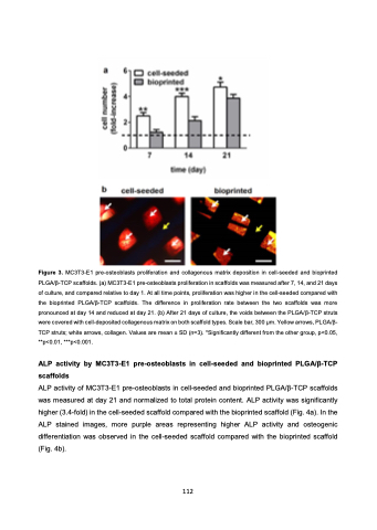
 Figure 3. MC3T3-E1 pre-osteoblasts proliferation and collagenous matrix deposition in cell-seeded and bioprinted PLGA/β-TCP scaffolds. (a) MC3T3-E1 pre-osteoblasts proliferation in scaffolds was measured after 7, 14, and 21 days of culture, and compared relative to day 1. At all time points, proliferation was higher in the cell-seeded compared with the bioprinted PLGA/β-TCP scaffolds. The difference in proliferation rate between the two scaffolds was more pronounced at day 14 and reduced at day 21. (b) After 21 days of culture, the voids between the PLGA/β-TCP struts were covered with cell-deposited collagenous matrix on both scaffold types. Scale bar, 300 μm. Yellow arrows, PLGA/β- TCP struts; white arrows, collagen. Values are mean ± SD (n=3). *Significantly different from the other group, p<0.05, **p<0.01, ***p<0.001.
ALP activity by MC3T3-E1 pre-osteoblasts in cell-seeded and bioprinted PLGA/β-TCP scaffolds
ALP activity of MC3T3-E1 pre-osteoblasts in cell-seeded and bioprinted PLGA/β-TCP scaffolds was measured at day 21 and normalized to total protein content. ALP activity was significantly higher (3.4-fold) in the cell-seeded scaffold compared with the bioprinted scaffold (Fig. 4a). In the ALP stained images, more purple areas representing higher ALP activity and osteogenic differentiation was observed in the cell-seeded scaffold compared with the bioprinted scaffold (Fig. 4b).
112
