
Page 93 - Fluorescence-guided cancer surgery
P. 93
Ex vivo imaging of resected lesion and sliced specimen
Ex vivo imaging of the resected specimens was performed at the Pathology department using the FLARE imaging system, which was previously described9. Images showed a uorescent signal in both lesions (Figure 2B). Of special note, the intensity of the uorescent signal decreased signi cantly over time, so a much weaker uorescence signal was measured during ex vivo imaging (i.e., T = 45 min after MB injection). The patchy uorescent signal of the main tumor was seen both in vivo and ex vivo. This was due to brous connective tissue lying over the paraganglioma. After bisection of the resected main lesion, a clear uorescent signal was seen throughout the tumor (Figure 2C).
Histopathology
Macroscopically, a nodular lesion was seen, yellow to black on cross section and surrounded by a thin capsule. The maximum diameter was 65 mm. Cranial from this lesion, a smaller lesion of 12 mm was seen, suspicious for a tumor- positive lymph node ( gure 2B). The excised tissue was xed in formalin, embedded in para n and tissue slides were stained with hematoxylin and eosin (HE). Immunohistochemical staining was also performed (Figure 4). Based on histopathological ndings, both lesions were diagnosed as paraganglioma with diameters of 65 and 12 mm, respectively.
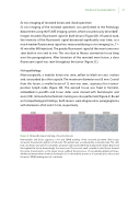
Figure 4. Histopathological staining of resected lesions
Hematoxylin and Eosin, Laguesse, S100 and SDHB staining of the resected specimen. Microscopy showed a characteristic pattern of cell nests. This pattern was accentuated by a reticulin stain. The cells had a moderate amount of eosinophilic cytoplasm and nuclei with nely clumped chromatin. Dispersed throughout the lesion, hemorrhagic foci were seen. The second, small, cranially located lesion showed the same characteristics as the larger lesion, without the presences of surrounding lymphoid tissue. Additional immunohistochemical staining for S100 showed the presence of sustentacular cells around the nests. SDHB staining was not conclusive.
Imaging of a paraganglioma 91
