
Page 92 - Fluorescence-guided cancer surgery
P. 92
90
Chapter 6
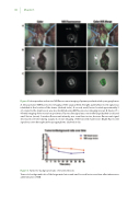
Figure 2. Intraoperative and ex vivo NIR uorescence imaging of primary and metastatic paragangliomas
A. Intraoperative NIR uorescence imaging of the surgical eld. A bright, patchy uorescent signal was identi ed at the location of the tumor (dashed circle). A second, small, lesion located approximately 5 cm cranial to the main lesion, was also identi ed using NIR uorescence imaging (arrow). B. Ex vivo (T = 45 min) imaging of the resection specimens. Fluorescent signal was seen in the large (dashed circle) and small lesion (arrow). A weaker uorescent intensity was seen than in vivo, because uorescent signal decreased over time during surgery. C. Ex vivo imaging of the bisected main lesion. Bright uorescent signal was seen throughout the paraganglioma (dashed circle).
Figure 3. Tumor-to-background ratio of resected lesions
Tumor-to-background ratio of the large main lesion and small second lesion over time after intravenous administration of MB.
