
Page 107 - Fluorescence-guided cancer surgery
P. 107
Intraoperative guidance in parathyroid surgery 105
that the resected tissue was parathyroid adenoma (N = 8) or hyperplastic parathyroid gland (N = 4). In 2 patients, the resected parathyroid adenoma did not uoresce. In summary, in 10 out of 12 patients with a proven parathyroid adenoma or hyperplastic parathyroid gland, the lesion was uorescent.
In 1 patient (#9), no adenoma was found during exploration of the neck. A thymectomy was performed and 6 non- uorescent lymph nodes were removed. During histological assessment of the resected tissue, no parathyroid adenomas were identi ed as well, although her intraoperative PTH assay showed a decrease from 25.0 to 1.4 pmol/L.
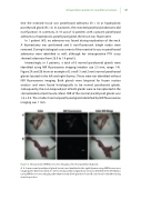
Interestingly, in 3 patients, a total of 6 normal parathyroid glands were identi ed using NIR uorescence imaging (median size 2.5 mm, range 1-4). Figure 2A and 2B show an example of 2 small (1 and 2 mm) normal parathyroid glands located in the left and right thymus. These were not identi ed without NIR uorescence imaging. Both glands were biopsied for frozen section analysis and were found histologically to be normal parathyroid glands. Subsequently, the non-biopsied part of both glands were re-transplanted in the sternocleidomastoid muscle. Mean SBR of the normal parathyroid glands was 1.8 ± 0.4. The smallest normal parathyroid gland identi ed by NIR uorescence imaging was 1 mm.
Figure 2. Intraoperative NIR uorescence imaging of normal parathyroid glands
A. A 2 mm normal parathyroid gland (arrow) was identi ed in the right thymus using NIR uorescence imaging after thymectomy. B. A 1 mm normal parathyroid gland (arrow) was identi ed in the left thymus using NIR uorescence imaging after thymectomy. Both glands in A and B could not be identi ed using visual inspection.
