
Page 106 - Fluorescence-guided cancer surgery
P. 106
104
Chapter 7
Table 2. Identi cation characteristics
of parathyroid adenomas
1 2 3 4 5
6 7 8 9 10 11 12
13
+ + + --- + - + +- + + + +
+ + + NP + -* + + +
- - +** --- --+
+NP+ +-+
+ 106
+ N/A
+ 102 and 132 + 113
+ 115;
109 and 134 + 34
+ 60
+ 87
+ 78 and 78 + N/A
+ 54
+ 61 and 145;
135 and 136 + 41
1.5
N/A
5.7 and 2.3
2.3
2.4 (adenoma);
1.4 and 2.0 (normal PTGs)
10.7
1.8 (ex vivo)
1.8
2.4 and 1.6 (normal PTGs)
N/A
1.7
3.8 and 1.7 (hyperplasia); 1.4 and 1.7 (normal PTGs)
2.2
Abbreviations: N/A, not applicable; NIRF, Near-infrared uorescence; NP, not performed, PTG, parathyroid gland; PTH parathyroid hormone; SBR, Signal-to-Background ratio; SPECT, single photon emission computed tomography.
* No in vivo NIRF detection of parathyroid adenoma. Ex vivo, the deepest located part of adenoma appeared to be uorescent. ** Identi cation of normal PTGs only using NIRF imaging.
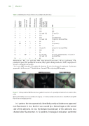
Figure 1. Intraoperative NIR uorescence-guided resection of a parathyroid adenoma located in the neck
During minimally-invasive parathyroid surgery, a 19 mm parathyroid adenoma is identi ed using NIR uorescence imaging (arrow).
In 1 patient, the intraoperatively identi ed parathyroid adenoma appeared non- uorescent in vivo, but this was caused by a hemorrhage on the ventral side of the adenoma. Ex vivo, the deepest located part of this adenoma also showed clear uorescence. In 12 patients, histological evaluation con rmed
Patient no.
Sestamibi- SPECT-CT
Preoperative ultrasound
NIRF detection
PTH decrease post-resection
Time between administration and NIRF detection (minutes)
SBR
