
Page 134 - Sentinel lymph node biopsy in oral cavity cancer - Inne J. den Toom
P. 134
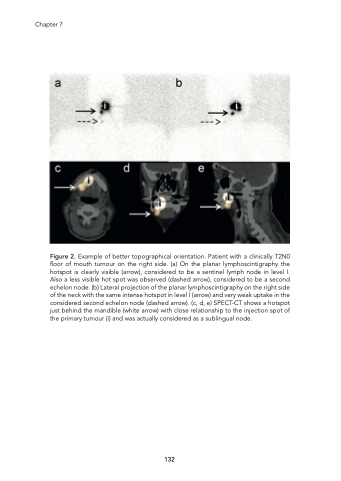
Chapter 7 Figure 2. Example of better topographical orientation. Patient with a clinically T2N0 floor of mouth tumour on the right side. (a) On the planar lymphoscintigraphy the hotspot is clearly visible (arrow), considered to be a sentinel lymph node in level I. Also a less visible hot spot was observed (dashed arrow), considered to be a second echelon node. (b) Lateral projection of the planar lymphoscintigraphy on the right side of the neck with the same intense hotspot in level I (arrow) and very weak uptake in the considered second echelon node (dashed arrow). (c, d, e) SPECT-CT shows a hotspot just behind the mandible (white arrow) with close relationship to the injection spot of the primary tumour (i) and was actually considered as a sublingual node. 132
