
Page 133 - Sentinel lymph node biopsy in oral cavity cancer - Inne J. den Toom
P. 133
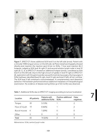
Figure 1. SPECT-CT shows additional SLN level II on the left side (arrow). Patient with a clinically T2N0 tongue tumour on the left side. (a) Planar lymphoscintigraphy showed directly post injection the injection spot (i) but no SLNs, 1 hour post injection (b) 2 hotspots, judged as SLN level IA right (1) and second echelon lymph node in level IV right (2). (c, d, e) SPECT-CT showed an additional hotspot (arrow), considered as SLN level II on the left side. Due to the high amount of uptake in level IV right on SPECT-CT (2), exploration with the gamma probe was performed during surgery. During surgery 3 SLNs had been identified (level IA right, level IV right and level II left), all hot, not blue. The SLN level II left contained a macrometastasis. A complementary neck dissection (selective I-IV) had been performed without additional metastasis on histopathological examination. No evidence of disease during follow-up of 32 months was observed. Table 1. Additional SLNs due to SPECT-CT imaging according to tumour localization 7 Location All patients Tongue 39 Floor of mouth 19 Buccal mucosa 4 Other 3 Total 65 Patients with Positive additional False additional SLNs SLNs negatives 5 (13%) 1 2 8 (42%) 1 2 1 (25%) 0 0 0 0 0 14 (22%) 2 4 Abbreviations: SLNs, sentinel lymph nodes 131
