
Page 89 - Cellular Imaging in Regenerative Medicine, Cancer and Osteoarthritis
P. 89
(~>10%) for acoustic pressures above 20 kPa PNP. Moreover, the percentage of SPIO positive cells increased up to ~12-15% with higher PNP for SPIO addition before the insonification. In contrast, SPIO addition at 5 and 15 min after ultrasound application resulted in much lower SPIO uptake (<8%). Similarly, the cell viability remained above 50% for all settings.
SPIO cell labeling
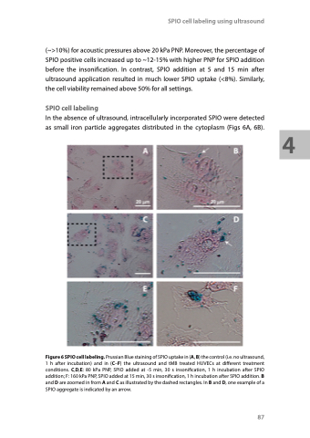
In the absence of ultrasound, intracellularly incorporated SPIO were detected as small iron particle aggregates distributed in the cytoplasm (Figs 6A, 6B).
Figure 6 SPIO cell labeling. Prussian Blue staining of SPIO uptake in (A, B) the control (i.e. no ultrasound, 1 h after incubation) and in (C–F) the ultrasound and tMB treated HUVECs at different treatment conditions. C,D,E: 80 kPa PNP, SPIO added at -5 min, 30 s insonification, 1 h incubation after SPIO addition; F: 160 kPa PNP, SPIO added at 15 min, 30 s insonification, 1 h incubation after SPIO addition. B and D are zoomed in from A and C as illustrated by the dashed rectangles. In B and D, one example of a SPIO aggregate is indicated by an arrow.
SPIO cell labeling using ultrasound
87
4
