
Page 88 - Cellular Imaging in Regenerative Medicine, Cancer and Osteoarthritis
P. 88
Chapter 4
Figure 4 The influence of SPIO incubation time on intracellular SPIO uptake efficiency. A SPIO positive cells. B Cell viability. HUVECs were treated with tMB and no ultrasound (- US) or ultrasound at varying PNP (40, 80, or 160 kPa) for 30 s. SPIO were added 5 min before insonification (-5 min). Incubation time after SPIO addition was varied from 5 min to 1 h and 3 h.
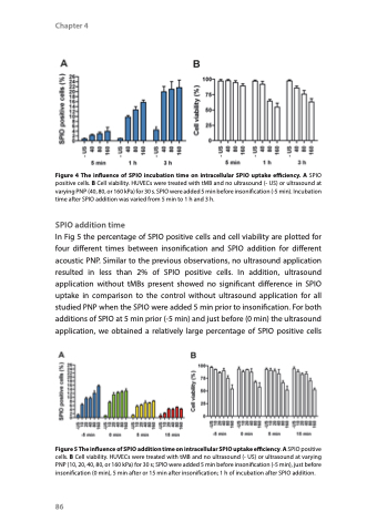
SPIO addition time
In Fig 5 the percentage of SPIO positive cells and cell viability are plotted for four different times between insonification and SPIO addition for different acoustic PNP. Similar to the previous observations, no ultrasound application resulted in less than 2% of SPIO positive cells. In addition, ultrasound application without tMBs present showed no significant difference in SPIO uptake in comparison to the control without ultrasound application for all studied PNP when the SPIO were added 5 min prior to insonification. For both additions of SPIO at 5 min prior (-5 min) and just before (0 min) the ultrasound application, we obtained a relatively large percentage of SPIO positive cells
Figure 5 The influence of SPIO addition time on intracellular SPIO uptake efficiency. A SPIO positive cells. B Cell viability. HUVECs were treated with tMB and no ultrasound (- US) or ultrasound at varying PNP (10, 20, 40, 80, or 160 kPa) for 30 s; SPIO were added 5 min before insonification (-5 min), just before insonification (0 min), 5 min after or 15 min after insonification; 1 h of incubation after SPIO addition.
86
