
Page 42 - Cellular Imaging in Regenerative Medicine, Cancer and Osteoarthritis
P. 42
Chapter 2
more dependent on the exact labeling protocol used. The highest intracellular iron loads without major adverse effects were obtained with incubation times of 24 h and intermediate labeling doses. Alternatively, better retention of label was observed after short incubation times and high labeling doses. This latter observation may be explained by differences in endocytosis kinetics, as depicted in Fig. 8. Labeling with high doses and short incubation times may result in large intracellular vesicles with multiple iron-oxide complexes. Labeling with low doses and long incubation times may result in small intracellular vesicles with just one iron-oxide complex. Following cell division the vesicles will be divided over the daughter cells; however, the vesicles themselves will not divide. Through these dynamics, the high dose and short
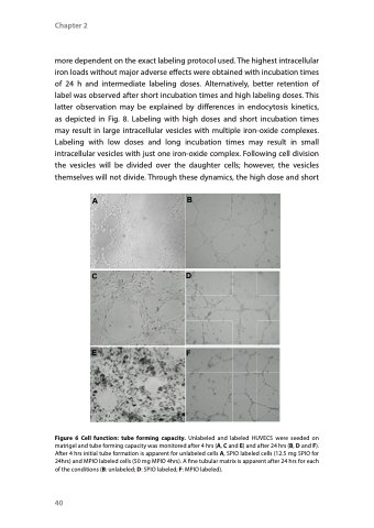
Figure 6 Cell function: tube forming capacity. Unlabeled and labeled HUVECS were seeded on matrigel and tube forming capacity was monitored after 4 hrs (A, C and E) and after 24 hrs (B, D and F). After 4 hrs initial tube formation is apparent for unlabeled cells A, SPIO labeled cells (12.5 mg SPIO for 24hrs) and MPIO labeled cells (50 mg MPIO 4hrs). A fine tubular matrix is apparent after 24 hrs for each of the conditions (B: unlabeled; D: SPIO labeled; F: MPIO labeled).
A
B
CD
EF
40
