
Page 18 - Cellular Imaging in Regenerative Medicine, Cancer and Osteoarthritis
P. 18
Chapter 1
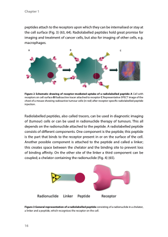
peptides attach to the receptors upon which they can be internalised or stay at the cell surface (Fig. 3) (63, 64). Radiolabelled peptides hold great promise for imaging and treatment of cancer cells, but also for imaging of other cells, e.g. macrophages.
Figure 2 Schematic drawing of receptor-mediated uptake of a radiolabelled peptide A Cell with receptors on cell surface B Radioactive tracer attached to receptor C Representative SPECT image of the chest of a mouse showing radioactive tumour cells (in red) after receptor-specific radiolabelled peptide injection.
Radiolabelled peptides, also called tracers, can be used in diagnostic imaging of (tumour) cells or can be used in radionuclide therapy of tumours. This all depends on the radionuclide attached to the peptide. A radiolabelled peptide consists of different components. One component is the peptide; this peptide is the part that binds to the receptor present in or on the surface of the cell. Another possible component is attached to the peptide and called a linker; this creates space between the chelator and the binding site to prevent loss of binding affinity. On the other site of the linker a third component can be coupled; a chelator containing the radionuclide (Fig. 4) (65).
Figure 3 General representation of a radiolabelled peptide consisting of a radionuclide in a chelator, a linker and a peptide, which recognises the receptor on the cell.
16
