
Page 17 - Cellular Imaging in Regenerative Medicine, Cancer and Osteoarthritis
P. 17
Introduction
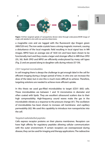
Figure 1 A Non targeted uptake of nanoparticles (brown dots) through endocytosis B MR image of single cells labelled in vitro with iron particles (black dots).
a magnetite core and are tagged with the fluorescent dye Dragon green (480/520 nm). The iron oxide crystals have a strong magnetic moment, causing a disturbance of the local magnetic field resulting in local signal loss in MR images. MPIO have an average size of 1630 nm and have been shown to be functionally inert and they create a larger and stronger effect in MRI than SPIO (55, 56). Both SPIO and MPIO are efficiently endocytosed by many cell types (Fig. 2) and are passed along to daughter cells during mitosis (57-59).
CD31 targeted microbubbles
In cell imaging there is always the challenge to get enough label in the cell for efficient imaging during a longer period of time. In vitro one can increase the dose of the label, but in vivo this is much more difficult to achieve. Therefore, targeting solutions are needed to achieve more efficient uptake.
In this thesis we used gas-filled microbubbles to target (CD31 (60)) cells. These microbubbles are between 1 and 10 micrometres in diameter and often coated with lipids. They are excellent ultrasound scatters due to their high compressibility. High-frequency sound waves make the gas in the microbubble vibrate as a response to the pressure change (61). The oscillation of microbubbles has been shown to increase cell membrane- and capillary permeability (62). We used this capability to introduce iron nanoparticles into the cell.
Targeted radiolabelled peptides
Cells express receptor proteins on their plasma membranes. Receptors can have high affinity for regulatory peptides allowing cellular communication with the outer environment. If certain receptors are overexpressed during disease, they can be used for imaging and therapy applications. The radioactive
15
1
