
Page 157 - Cellular Imaging in Regenerative Medicine, Cancer and Osteoarthritis
P. 157
SPECT imaging of pro-inflammatory macrophages
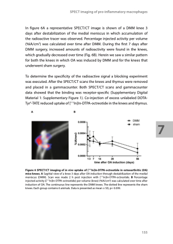
In figure 6A a representative SPECT/CT image is shown of a DMM knee 3 days after destabilization of the medial meniscus in which accumulation of the radioactive tracer was observed. Percentage injected activity per volume (%IA/cm3) was calculated over time after DMM. During the first 7 days after DMM surgery, increased amounts of radioactivity were found in the knees, which gradually decreased over time (Fig. 6B). Herein we saw a similar pattern for both the knees in which OA was induced by DMM and for the knees that underwent sham surgery.
To determine the specificity of the radioactive signal a blocking experiment was executed. After the SPECT/CT scans the knees and thymus were removed and placed in a gammacounter. Both SPECT/CT scans and gammacounter data showed that the binding was receptor-specific (Supplementary Digital Material 1: Supplementary Figure 1). Co-injection of excess unlabeled DOTA- Tyr3-TATE reduced uptake of [111In]In-DTPA-octreotide in the knees and thymus.
Figure 6 SPECT/CT imaging of in vivo uptake of [111In]In-DTPA-octreotide in osteoarthritic (OA) mice knees. A Sagittal view of a knee 3 days after OA induction through destabilization of the medial meniscus (DMM). Scan was made 2 h post injection with [111In]In-DTPA-octreotide. B Percentage injected activity ([111In]In-DTPA-octreotide) per volume (knee) (%IA/cm3) was calculated over time after induction of OA. The continuous line represents the DMM knees. The dotted line represents the sham knees. Each group contains 6 animals. Data is presented as mean ± SD, p= 0.099.
155
7
