
Page 153 - Cellular Imaging in Regenerative Medicine, Cancer and Osteoarthritis
P. 153
SPECT imaging of pro-inflammatory macrophages
Results
Somatostatin receptor subtype 2 mRNA expression
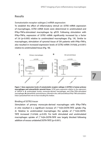
To establish the effect of inflammatory stimuli on SSTR2 mRNA expression of macrophages, SSTR2 mRNA levels were determined in unstimulated and IFNγ+TNFα-stimulated macrophages by qPCR. Following stimulation with IFNγ+TNFα, expression of SSTR2 mRNA significantly increased by a factor of 3.6 (p<0.005) relative to unstimulated macrophages (Fig. 1A). Similar to macrophages, stimulation of synovial tissue of OA patients with IFNγ+TNFα, also resulted in increased expression levels of SSTR2 mRNA (10-fold, p<0.001) relative to unstimulated tissue (Fig. 1B).
Figure 1 Gene expression levels of somatostatin receptor subtype 2 (SSTR2) in human primary macrophages and osteoarthritic synovial tissue. SSTR2 gene expression relative to the expression of GAPDH in A unstimulated human monocyte-derived macrophages and IFNγ+TNFα-stimulated macrophages. B osteoarthritic synovial tissue, cultured in vitro, with and without IFNγ+TNFα stimulation. Data is presented from min to max including the median.
Binding of SSTR2 tracer
Stimulation of primary monocyte-derived macrophages with IFNγ+TNFα in vitro resulted in a significant increase of [111In]In-DOTA-TATE uptake (Fig. 2). Relative to unstimulated macrophages the uptake of [111In]In-DOTA- TATE increased (1.6-fold, p<0.05). For both stimulated and unstimulated macrophages uptake of [111In]In-DOTA-TATE was largely blocked following addition of excess unlabeled DOTA-TATE (p<0.001).
151
7
