
Page 98 - Assessing right ventricular function and the pulmonary circulation in pulmonary hypertension Onno Anthonius Spruijt
P. 98
6
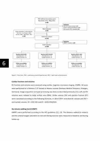
Figure 1: Flow chart. PAH = pulmonary arterial hypertension; RHC = right heart catheterization.
Cardiac function and volumes
RV function and volumes were assessed using cardiac magnetic resonance imaging (CMRI). All scans were performed on a Siemens 1.5T Sonata or Avanto scanner (Siemens Medical Solutions, Erlangen, Germany). Image acquisition and post-processing was done as described previously [11]. Left and RV volumes were indexed to body surface area (BSA). Stroke volume (SV) and ejection fraction (EF) were calculated according to the following formulas, in which EDV= end-diastolic volume and ESV = end-systolic volume: SV = EDV-ESV and EF = (EDV-ESV)/EDV.
Six-minute walking test (6 MWT)
6MWTs were performed according to the ATS guidelines [12, 13]. The distance walked (in meters) and the arterial oxygen saturation at rest and during exercise were measured at baseline and during follow-up.
