
Page 84 - Tailoring Electrospinning Techniques for Regenerative Medicine - Marc Simonet
P. 84
CHAPTER 4
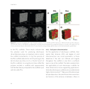
Figure 4.2 Computer tomography (CT) scans of typical examples of conv. (A & B) and LTE (C & D) spun sca olds all with 9 μm fiber diameter, spun from PCL (A & C) or PLA (B & D) over 60 minutes. A 3 dimensional rendering of the spatial fiber distances is shown on the right (a-d). The sections showed are taken in the middle of the CT scans from the corresponding sca olds shown on the le , labeled with capital letters.
to the PCL sca olds. These results indicate that the polymer used for producing electrospun 3D-architectures plays an important role to control the sca old characteristics. In fact the porosity of the PLA sca olds a er 60 min of LTE spinning on the 50mmdrumwas99.5±0.3%vs.the98.7±0.5%of the PCL sca olds. So using the 8.4 times sti er PLA polymer resulted in sca olds with approximately half the fiber density compared to the sca olds spun with the so er PCL.
4.4.2 Void space characterization
For the application of electrospun sca olds, their spatial fiber distance (SFD) and degree of void interconnections are crucial parameters, as these determine if the cells can infiltrate and migrate throughout the sca old or only form a confluent layer on top of the sca old. The latter severely limits the applicability of such electrospun sca olds for three dimensional tissue engineering applications. Generally, electrospun sca olds are considered to be highly interconnected.12,42 Because of the dense layer by layer deposition, the size of these interconnections are fractions of the distance in-between 2 fibers in
82
