
Page 134 - Tailoring Electrospinning Techniques for Regenerative Medicine - Marc Simonet
P. 134
CHAPTER 6
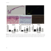
Figure 6.6 Histology (a) HE stained cells infiltrate the entire sca old (b) PSR staining for collagen, with the collagen formation near the reinforcement ring. (c) HE staining cells lining the sca old and cells infiltrate the sca old. (d) aSMA staining for myofibrolasts in red (e) PSR staining for collagen. (f) Number of cells in the sca old per 20x magnification including cells lining the sca old and cells infiltrating the sca old. (g) Number of cells infiltrated the sca old per hpf (40x magnification) (e) Mean amount of collagen. Statistically no di erences were found.
132
