
Page 133 - Tailoring Electrospinning Techniques for Regenerative Medicine - Marc Simonet
P. 133
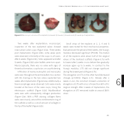
Figure 6.5 Macroscopic outcome of the implants (a) 2 weeks (b) 3 weeks (c) 4 weeks and (d) 5 weeks post implantation.
Two weeks a er implantation, macroscopic inspection of the two explanted valves showed intact pliant valve cusps (Figure 6.5a). Three weeks post implantation (Figure 6.5b), some weak spots were observed in the belly of the cusps in all cases. A er 4 weeks (Figure 6.5c), holes appeared and a er 5 weeks (Figure 6.5d) valve leaflets were torn apart. Macroscopically, there was no valve with signs of thrombus formation, cusp fusion, or cusp thickening.
Infiltration of mainly neutrophils and monocytes were seen throughout the entire leaflet cross section with HE staining on the two valves explanted two weeks a er implantation (Figure 6.6a). Additionally a few macrophages were observed. Cells were mainly located at the basis of the valve cusps, lining the electrospun sca old (Figure 6.6d). Myofibroblast were seen with subsequently collagen production (Figure 6.6e). With a PSR staining, collagen fibers were seen mainly around the reinforcement ring of the sca old as well as a small amount of collagen in the tip of the leaflet (Figure 6.6b).
Small strips of the explants at 2, 3, 4 and 5 weeks were tested for their mechanical properties. Evaluated over the period of the 5 weeks, the Young’s modulus decreased significant (P<0.05). The moduli of all the explants were about half of the original values of the sterilized sca olds (Figure 6.7a) with its lowest a er 3 weeks in-vivo before then gradually increase again up to 5 weeks. In contrast to the Young’s modulus, UTS did not change significant during the 5 week period (Figure 6.7b).
The elongation at UTS on the other hand decreased 6 strongly (p<0.0001) (Figure 6.7c). Already a er 2
weeks in-vivo, the construct showed a reduction of
elongation at UTS with a loss of more than 50% of its
original strength. A er 3 weeks of implantation, the elongation at UTS remained stable at around 150% strain.
FROM A POLYMER TOWARDS AN IN-SITU TISSUE ENGINEERED HEART VALVE
131
