
Page 105 - Tailoring Electrospinning Techniques for Regenerative Medicine - Marc Simonet
P. 105
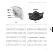
Figure 5.3 (a) The configuration of the fibrosa, spongiosa and ventricularis within the aortic valve leaflet (adapted from Mol et al.28). (b) Typical collagen fiber structure in an aortic valve leaflets (adapted from Sauren29). The commissures are denoted by ‘c’.
very robust tissues, serving a life-dependent function. Microscopically, the leaflets consist of three function-tailored tissue layers, maintained by the valvular interstitial cells, covered by endothelium. The fibrosa layer at the arterial side of the leaflets is mainly composed of circumferentially aligned collagen fibers. This layer is considered the main load-bearing layer of the leaflet and prevents excessive stretching during diastole. The ventricularis layer at the outflow surface of the leaflets mainly consists of radially aligned elastin. The elastin in this layer is believed to restore the collagen fiber geometry to its original corrugated geometry during systole allowing it to be stretched again in the next heart cycle. In between the fibrosa and the ventricularis, a loose spongiosa
layer is present, containing an abundant amount of 5 proteoglycans. This layer is suggested to serve as a
shock absorber upon closure of the valve and enables
sliding of the individual layers for optimal functioning.
The typical layering of the aortic valve leaflet and the collagen fiber structure in the fibrosa of the aortic valve leaflet is demonstrated in Figure 5.3.
6.4.3 Cell-matrix interactions in semilunar heart valves
During development, heart valve formation goes through coordinated sequences of growth, di erentiation and organization that are orchestrated by spatial and temporal gradients of multiple regulatory factors. Accumulating evidence suggests
ELECTROSPINNING FOR HEART VALVE TISSUE REGENERATION
103
