
Page 103 - Tailoring Electrospinning Techniques for Regenerative Medicine - Marc Simonet
P. 103
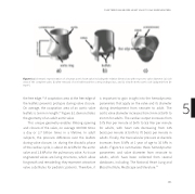
Figure 5.1 Schematic representation of a human aortic heart valve including the relative dimensions with respect to valve diameter: (a) side view of the complete valve, (b) a er removal of one leaflet and the corresponding sinus, and (c) view from the aortic side (adapted from De Hart25).
the free edge.24 A coaptation area at the free edge of the leaflets prevents prolapse during valve closure. On average, the coaptation area of an aortic valve leaflets is 5 mm in length.24 Figure 5.1 demonstrates the geometry of an adult aortic valve.
This unique geometry enables lifelong opening and closure of the valve, on average 100’000 times a day or 3.7 billion times in a lifetime. In adult subjects, the pressure di erence over the leaflets during valve closure, i.e. during the diastolic phase of the cardiac cycle, is about 10-12 kPa for the aortic valve and 1.5 kPa for the pulmonary valve. As tissue engineered valves are living structures, which allow for growth and remodeling, they represent attractive valve substitutes for pediatric patients. Therefore, it
is important to gain insight into the hemodynamic 5 parameters that apply on the valve and its diameter
during development from neonate to adult. The
aortic valve diameter increases from 9 mm at birth to
23 mm for adults. The cardiac output increases from 0.75 liter per minute at birth to 5.5 liter per minute for adults, with heart rate decreasing from 145 beats per minute at birth to 70 beats per minute in adults. Finally, the transvalvular pressure at diastole increases from 5 kPa at 1 year of age to 10 kPa in adults. Figure 5.2 summarizes these hemodynamic parameters and valve diameter from neonate to adults, which have been collected from several databases, including, The National Heart Lung and Blood Institute, MedScape and literature.23
ELECTROSPINNING FOR HEART VALVE TISSUE REGENERATION
101
