
Page 99 - scheppingen
P. 99
DYSREGULATION OF THE (IMMUNO)PROTEASOME PATHWAY IN MCD
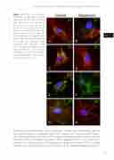
Figure 8 Effects of rapamycin treatment on proteasome subunit expression in FCD type II derived cells. Expression of β1 (A and B; green), β1i (C and D; green), β5 (E and F; red) and β5i (G and H; green) in untreated FCD cells (left panels) and in FCD cells after 48 hours treatment with 100 nM rapa- mycin (right panels). Expression of all subunits was decreased after treatment with rapamycin. Cells were counterstained with phalloi- din (actin filaments; red in A-D and G-H, green in E and F) and diamid- ino-2-phenylindole, DAPI (nuclei; blue). Scale bar in A: 15 μm.
including immunoproteasome subunit expression. Accordingly, we observed a positive correlation between immunoproteasome subunit expression in neurons and pS6 expres- sion, indicating the activation of the mTOR signal transduction pathway. The relationship between mTOR and proteasome system is also supported by the in vitro experiments showing that inhibition of the mTOR pathway by the potent allosteric mTORC1 inhibitor rapamycin was able to reduce the level of expression of inducible proteasome subunits
97
four
