
Page 34 - scheppingen
P. 34
two
ly-monthly), antiepileptic drug (AED) management at surgery, type of epilepsy surgery, duration of active epilepsy, last available postsurgical seizure outcome (according to Engel’s score), average intelligence quotient (IQ) with global cognitive performance and presence/absence of autism 12.
Interobserver agreement
For case evaluation, six different neuropathologists gained access to an online virtual slide system (Digital Slidebox 4.5, Slidepath; Leica Microsystems, Dublin, Ireland). These reviewers were asked to classify a subset of ten randomly selected cases provided with three basic stainings (H&E, SMI32 and vimentin) according to the novel scheme of pat- terns discussed in this manuscript. After 21 days the platform was closed. None of the reviewers had access to the results of the others.
Statistical analysis
Statistical analysis was performed on SPSS 21 (IBM, PASW Statistics, USA). The distribu- tions of pS6, SMI32 and vimentin were left skewed. Therefore, the data were log-trans- formed prior to statistical analysis. Hierarchical clustering (Ward’s method with squared Euclidian distances) and one-way ANOVA were used for specify the tuber patterns (S2 Fig). Due to the lack of normality and non-equality of variances non-parametric testing (independent-sample Kruskal-Wallis test) as well as Kendall-tau correlation were used to analyse the data. Partial correlation was applied if data needed to be corrected for another variable. The Chi-squared test was applied for analysing categorical data. The κ coefficient was calculated to address inter-rater variability. In our study κ was inter- preted as follows: <0.2, poor agreement; 0.2–<0.4, fair agreement; 0.4–<0.6, moderate agreement; 0.6–<0.8, good agreement; 0.8–1.0, very good agreement. Bootstrapping was
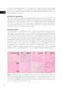
Figure 1 Histology of tuber variants. A. Perilesional cortex (Cx) and white matter (WM) of an 8-year old male patient. B. Type A tuber (Cx and WM) in a 5-year old female patient with a tuber in the frontal lobe. C. Type B tuber (Cx and WM) located in the parietal lobe of an 8-year old boy (same as in Fig. 1A). D. Type C tuber of a 2-year old male patient located in the frontal lobe. Scale bar in D = 100μm and applies also to A, B and C.
32
