
Page 18 - scheppingen
P. 18
one
16
children, always coincide with high levels of proinflammatory mediators 98. Furthermore, upregulation of inflammatory mediators has been found in different human epilepsies, irrespective of the underlying pathologic mechanism or cause 94, 99, 100. This indicates that brain inflammation might be intrinsic to some epileptic pathologies, rather than being just a consequence of seizure activity.
Traditionally, the brain was considered as an immune privileged organ due to the presence of the blood-brain barrier (BBB), which limits the trafficking of peripheral immune cells, and the lack of a draining lymphatic system. However, all cell types present the brain are involved in the classical innate immune response, and together with periph- eral leukocytes that infiltrate the brain upon inflammatory challenges accounting for the adaptive immune response, a CNS immune response can be evoked in response to injury, pathogens or other pathological events 94-96. Inflammation in the brain coincides with the activation of several inflammatory cascades, accompanied by the production of proinflammatory mediators by different cells in the brain. Amongst these proinflamma- tory factors are cytokines that include interleukins, tumor necrosis factors (TNFs), inter- ferons (IFNs), and growth factors (like transforming growth factor-β, TGF-β), but also chemokines, prostaglandins and complement factors 97, 101, 102. These proinflammatory mediators can induce leakage of the BBB leading to albumin extravasation in the brain, which is correlated with an increased number of seizures and progression of epilepsy 103. Furthermore, these factors induce transcriptional changes in glutamate and gamma-am- inobutyric acid (GABA) receptors, increased glutamate release, neurogenesis, sprouting and angiogenesis, all together leading to increased excitability and development of epi- lepsy 94-97. Additionally, high mobility group box 1 protein (HMGB1), is released as ‘danger signal’ upon inflammation, targeting Toll-like receptor 4 (TLR4) signaling to promote seizure activity. This, on its turn, induces further release of HMGB1, resulting in a positive feedback loop reinforcing seizures and brain inflammation 104.
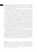
Inflammation in the brain involves both glial cells and infiltrating leukocytes, the white blood cells of the adaptive immune system that are responsible for the protection against foreign pathogens. Glial cells are important regulators of various inflammatory processes in the brain. CNS glia include astrocytes, NG2-glia or oligodendrocyte progen- itor cells, oligodendrocytes and microglia. Both microglia and astrocytes are found to be prominently reactive, called microgliosis and astrogliosis, respectively, in various epileptic pathologies 105. Microgliosis and astrogliosis lead to the release of proinflammatory sig- naling molecules that are able to alter connections between glial cells and neurons, lead- ing to enhanced excitability which contributes to epileptogenesis (see Fig 4) 96, 100, 106-108.
Figure 4. Schematic overview depicting mechanisms contributing to epileptogenesis in TSC. A better understanding of these mechanisms may guide the recognition of novel targets for precision medicine approaches to TSC-related epilepsy based on targeted combination thera- pies. TLR4: toll-like receptor 4; IL-1β: interleukin-1β; RAGE: receptors for advanced glycation end products; cation chloride cotransporters: NKCC1 and KCC2; mGluRs: metabotropic glutamate receptors; iGluRs: ionotropic glutamate receptors; Glu: glutamate; GABAAR: gamma-aminobutyric acid type A receptor; BBB: blood-brain barrier.
