
Page 15 - scheppingen
P. 15
GENERAL INTRODUCTION & OUTLINE OF THE THESIS
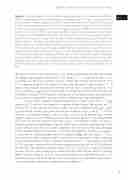
Figure 2. Simplified diagram of the mTORC1 signaling pathway and its downstream effectors. mTORC1 signaling plays a central role during neural development. TSC1 and TSC2 proteins form a heterodimeric complex, together with TBC1 domain family member 7 (TBC1D7), that functions as a GTPase-activating protein (GAP) for small G-protein Ras homology enriched in brain (Rheb), acting as an upstream regulator of the mTOR signaling pathway. mTORC1 consists of five com- ponents, including: mTOR, the two positive regulatory subunits rapamycin-associated protein of TOR (Raptor) and mammalian lethal with SEC13 protein 8 (mLST8), and two negative regulators proline-rich Akt substrate of 40 kDa (PRAS40) and DEP domain-containing mTOR-interacting protein (Deptor). Rapalogs (rapamycin derivatives; such as everolimus and sirolimus) are po- werful inhibitors of mTORC1 activity. PI3K: PI3kinase; PDK1: phosphoinositide-dependent kinase-1; PTEN: Phosphatase and tensin homologue; AMPK: AMP-activated protein kinase; TBC1D7: TBC1 domain family member 7; LKB1: tumor suppressor liver kinase B1; STRADα: STE20-related kina- se adaptor alpha; Rheb: ras homolog enriched in brain; mTORC: mammalian target of rapamycin complex; DEPDC5: DEP Domain Containing 5; NPRL3: NPR3 Like, GATOR1 Complex Subunit; FKBP: FK506 binding protein. HIF1 alpha: Hypoxia-inducible factor 1-alpha; VEGF: Vascular endothelial growth factor; S6K1: p70S6kinase; S6: ribosomal S6 protein; eIF4E: binding of eukaryotic transla- tion.
focal dyslamination, isolated giant cells), that may contribute to the complex and variable neurological phenotype encountered in TSC patients 38, 51-54. Lesion formation in TSC may follow the classical mutational ‘‘two-hit’’ model with somatic inactivation of TSC1/ TSC2. However, second hit mutations are not always detected in cortical tubers 55, 56. A recent study indicates that only one-third of cortical tubers are driven by somatic TSC1/ TSC2 inactivation, suggesting that either only a small portion of cells within the tuber are affected by a second hit, limiting their identification (mutational burden below detection limits) or that a mono-allelic mutation could be sufficient for tuber development 35.
Cortical tubers represent focal developmental malformations that, in a large majority of TSC patients, are detected as single or multiple lesions (for reviews see 26, 39). They consist in areas of cortical dyslamination that contain different cell types, includ- ing dysmorphic neurons, giant cells and reactive astrocytes (Fig 1, 3 and 4) 37, 39. Cortical layering is markedly disturbed in cortical tubers and analysis of cortical layer markers suggests a dysmaturation affecting early and late migratory patterns, with a more severe impairment of the late stage of maturation (Fig 3) 57. Dysmorphic neurons are charac- terized by abnormal morphology, abnormal orientation and abnormally large sizes. They express different cortical layer markers, regardless of their laminar location and display an immunophenotype that resembles that of cortical projection neurons and suggests an alteration of a selected population of intermediate progenitor cells (Fig 3) 57, 58. Giant cells have been shown to express both neuronal and immature glial markers, indicating a failure to differentiate prior to migration into the cortex 59, 60. Enhanced activation of mTOR signaling is evidenced by enhanced phospho-activation of the mTOR effectors p70S6kinase and ribosomal S6 protein (pS6, see Fig 1 G and Fig 2) in both dysmorphic neurons and giant cells, providing the molecular explanation for their aberrant morpho- logical features. However, pS6 has been also detected immunohistochemically in the perituberal cortex and activation of mTOR signaling pathway, evidenced by pS6 expres- sion, has also been reported in acquired forms of epilepsy 61-65.
13
one
