
Page 56 - Imaging of Osteoarthritis and Rheumatoid Arthritis in Hand Joints
P. 56
Chapter 3
Table 2. Presence and absence of detected CMC1 OA by modality for both readers.
CMC1 OA Reader 1 CR positive
CR negative
CT positive
25 3
CT negative
2 0
CMC1 OA Reader 2 CR positive
CR negative
CT positive
25 3
CT negative
0 2
CT negative
1 13
CR = conventional radiography, CT = computed tomography
Table 3. Presence and absence of detected STT OA by modality for both readers.
STT OA Reader 1 CR positive
CR negative
CT positive
6 12
CT negative
0 11
STT OA Reader 2 CR positive
CR negative
CT positive
10 5
CR = conventional radiography, CT = computed tomography
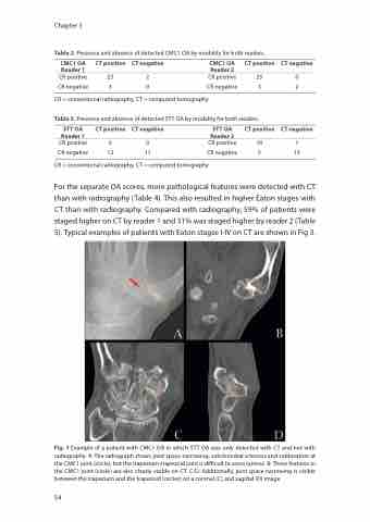
For the separate OA scores, more pathological features were detected with CT than with radiography (Table 4). This also resulted in higher Eaton stages with CT than with radiography. Compared with radiography, 59% of patients were staged higher on CT by reader 1 and 31% was staged higher by reader 2 (Table 5). Typical examples of patients with Eaton stagse I-IV on CT are shown in Fig 3.
Fig. 1 Example of a patient with CMC1 OA in which STT OA was only detected with CT and not with radiography. A: The radiograph shows joint space narrowing, subchondral sclerosis and subluxation at the CMC1 joint (circle), but the trapezium-trapezoid joint is difficult to asses (arrow). B: These features in the CMC1 joint (circle) are also clearly visible on CT. C-D: Additionally, joint space narrowing is visible between the trapezium and the trapezoid (circles) on a coronal (C) and sagittal (D) image
54
