
Page 193 - Demo
P. 193
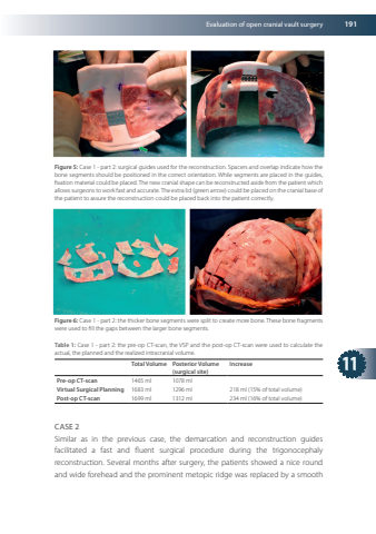
Evaluationofopencranialvaultsurgery19111Figure5Case1-part2surgicalguidesusedforthereconstructionSpacersandoverlapindicatehowthebonesegmentsshouldbepositionedinthecorrectorientationWhilesegmentsareplacedintheguidesfixationmaterialcouldbeplaced Thenewcranialshapecanbereconstructedasidefromthepatientwhichallowssurgeonstowork fastandaccurate Theextralid(greenarrow)couldbeplacedonthecranialbaseofthepatienttoassurethereconstructioncouldbeplacedbackintothepatientcorrectlyFigure6Case1-part2thethickerbonesegmentsweresplittocreatemorebone ThesebonefragmentswereusedtofillthegapsbetweenthelargerbonesegmentsTable1Case1-part2thepre-opCT-scantheVSPandthepost-opCT-scanwereusedtocalculatetheactualtheplannedandtherealizedintracranialvolumeTotal VolumePosterior Volume(surgicalsite)IncreasePre-opCT-scan1465ml1078mlVirtualSurgicalPlanning1683ml1296ml218ml(15%oftotalvolume)Post-opCT-scan1699ml1312ml234ml(16%oftotalvolume)CASE2SimilarasinthepreviouscasethedemarcationandreconstructionguidesfacilitatedafastandfluentsurgicalprocedureduringthetrigonocephalyreconstructionSeveralmonthsaftersurgerythepatientsshowedaniceroundandwideforeheadandtheprominentmetopicridgewasreplacedbyasmooth
