
Page 165 - Demo
P. 165
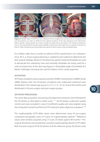
Longitudinal evaluation16310Figure2NotablestepsinOCVRfromourinstitutionPriortosurgerythereconstructionisvirtuallyplannedandexecutedbyourteamtodeterminetheoptimalpositionsoftheskullsegmentsandosteotomiesPatientspecificsurgicalguidesareusedtotransfertheplanningtothepatientLeftdrawingosteotomylinesontheexposedskullusingtheguidesMiddlepositioningofskullsegmentsinaspecificmoldpriortofixationRightrepositioningofexpandedskullsegmentfixedinplaceusingresorbableplatesForchildrenolderthan6monthsatreferralOCVRisperformedinourinstitutionSince2013avirtualsurgicalplanningiscreatedforeachpatienttodeterminethebestsurgicalstrategyBasedonthisplanningspecificdrawingtemplatesareusedtodemarcatetheosteotomylinesandassemblytemplatesarebeingusedforaswiftreconstructionoftheskullcap(Figure2)Resorbableplates(SonicWeldKLSMartin TuttlingenGermany)areusedforfixationofthecranialsegments3DPHOTOS3DPhotos ofpatients were acquiredusing the 3DMD Cranial System(3dMDCranial3dMDAtlantaUSA)The3Dphotosofpatientswhounderwenttreatmentweredistributedinthenearestagegroup(3691215182436and48months)anddistributedinthepre-surgeryandpost-surgerygroups3DPHOTOPROCESSINGThesamedataacquisitionprocessingandalignmentprotocolswerefollowedforthe3Dphotosasdescribedinearlierwork15–17All3Dphotosunderwentqualitycontrolandwereexcludedincaseofinsufficientqualityandwerealignedusingthe Computed CranialFocalPoint(CCFP)method to allowobjectiveevaluation1618ThescaphocephalyCCFP-offsetvaluesweredeterminedusing20pre-surgerycomputedtomographyscans(CT-scans)ofscaphocephalypatients16ReferencevaluesweresimilarlyacquiredusingCT-scansofinfantsaged0-48months15Presurgical3Dphotos werepositionedusing thescaphocephaly-specificCCFP-offsetBoththepost-surgicalOCVR3Dphotosandthereferencegroup3Dphotoswere
