
Page 142 - Demo
P. 142
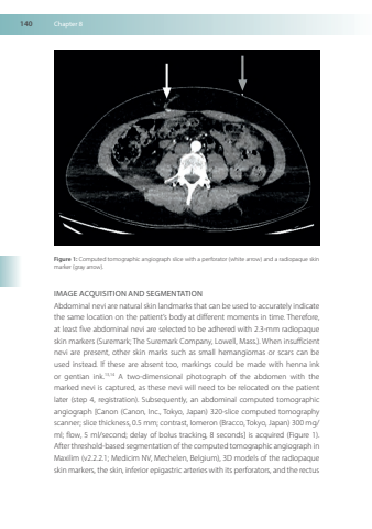
140Chapter8Figure1Computedtomographicangiographslicewithaperforator(whitearrow)andaradiopaqueskinmarker(grayarrow)IMAGEACQUISITIONANDSEGMENTATIONAbdominalneviarenaturalskinlandmarksthatcanbeusedtoaccuratelyindicatethesamelocationonthepatient’sbodyatdifferentmomentsintime Thereforeatleastfiveabdominalneviareselectedtobeadheredwith23-mmradiopaqueskinmarkers(Suremark; TheSuremarkCompanyLowellMass)WheninsufficientneviarepresentotherskinmarkssuchassmallhemangiomasorscarscanbeusedinsteadIftheseareabsenttoomarkingscouldbemadewithhennainkorgentianink1314Atwo-dimensionalphotographoftheabdomenwiththemarkedneviiscapturedastheseneviwillneedtoberelocatedonthepatientlater(step4registration)SubsequentlyanabdominalcomputedtomographicangiographCanon(CanonIncTokyoJapan)320-slicecomputedtomographyscanner;slicethickness05mm;contrastIomeron(Bracco TokyoJapan)300mg/ml;flow5ml/second;delayofbolustracking8secondsisacquired(Figure1)After threshold-basedsegmentationof thecomputed tomographic angiographinMaxilim(v2221;MedicimNV,MechelenBelgium)3Dmodelsoftheradiopaqueskinmarkers theskininferiorepigastricarteries withitsperforatorsand therectus
