
Page 120 - New epidemiological and PSMA-expression based paradigms in salivary gland tumors
P. 120
Chapter 7
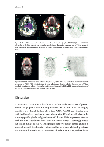
Figure 3: Case B. Transverse slices of radiotherapy dose distribution (A), fused PET/CT (B) and PSMA PET (C) at the level of the parotid and retropharyngeal glands, illustrating complete loss of PSMA uptake in pharyngeal wall glands and in the deep lobe of the left parotid gland (green arrows), which received a high dose.
Figure 4: Case C. Transverse slice of fused PET/CT (A), PSMA PET (B), and lateral maximum intensity projection of PSMA PET with indication of prior radiotherapy field in red (C). Complete loss of PSMA uptake is seen in most salivary glands after radiotherapy. Remarkably, PSMA PET indicates hypertrophy of the spared minor salivary glands in the lips (green arrows).
Discussion
In addition to the familiar role of PSMA PET/CT in the assessment of prostate cancer, we propose a new and very different use for this molecular imaging modality. Our clinical findings show that PSMA PET/CT can visualise areas with healthy salivary and seromucous glands after RT, and identify damage by showing specific glands and gland areas with loss of PSMA expression coherent with the dose distribution from prior RT. PSMA PET/CT seemingly detects subclinical damage in case A. The signal gradient over the left parotid gland is in concordance with the dose distribution, and has an inverse relationship between the treatment dose and tracer accumulation. This also indicates a spatial resolution
118
