
Page 118 - New epidemiological and PSMA-expression based paradigms in salivary gland tumors
P. 118
116
Chapter 7
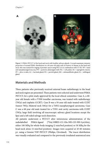
Figure 1: PSMA PET/CT of the head and neck with healthy salivary glands. Coronal maximum intensity projection of normal PSMA distribution in a 60-year-old male with no history of disease in the head and neck, who was scanned for staging of prostate cancer using PET (A) in combination with CT (B). NP = nasal and nasopharyngeal mucosa; PW = posterior pharyngeal wall; PAL = soft palate; SG = supraglottic larynx; PV = plica vocalis; LG = lacrimal gland; PG = parotid gland; SM = submandibular gland; SL = sublingual gland.
Materials and Methods
Three patients who previously received external beam radiotherapy to the head and neck region are presented. These patients were selected and underwent PSMA PET/CT, for a pilot study approved by the local ethical committee. Case A, a 60- year old female with a T3N0 tonsillar carcinoma, was treated with radiotherapy (70Gy) and cisplatin (CCRT). Case B was a 50-year old male treated with CCRT (tumor 70Gy; bilateral neck 54Gy) for a T4N2 nasopharyngeal carcinoma. Case C was a 68-year old male treated for a T3N1 oral cavity carcinoma with CCRT (70Gy, large field involving all macroscopic salivary gland locations except the lips) and a left sided salvage neck dissection.
All patients underwent a PET/CT after intravenous administration of the radiolabelled PSMA-ligand [68Ga]-HBED-CC-Glu-NH-CO-NH-Lys(Ahx), either 100 MBq for whole-body imaging (2 min/bed position) or 50 MBq for the head-neck alone (4 min/bed position). Images were acquired at 45-60 minutes p.i. using a Gemini TOF PET/CT (Philips, Cleveland). The tracer distribution was visually evaluated and compared to the previously irradiated anatomical area
