
Page 97 - Synthesis of Functional Nanoparticles Using an Atmospheric Pressure Microplasma Process - LiangLiang Lin
P. 97
Solvent-Free Nickel Nanoparticles Synthesis and Engineering ‒Controllable Magnetic Properties
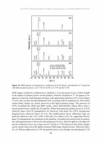
Figure 5.4 XRD patterns of nanoparticles synthesized at fixed Ni(cp)2 concentration of 35 ppm but with different plasma powers: (a) 1.3 W, (b) 2.0 W, (c) 2.7 W and (d) 3.4 W.
TEM images of particles synthesized at condition 1-4 are presented to give a better insight of the impact of plasma power on the products. Particles prepared at 1.3 W appear to be spherical, relatively small and quite uniform, with approximate diameters of 5-15 nm (Figure 5.5 a-b). One can also note that abundant CNTs coexisting with Ni nanoparticles, from which carbon lattice fringes are clearly observed in the high-resolution image. The presence of CNTs reconfirms the SEM and XRD results, where fibril/felt-like carbon fibers form a closely packed layer outside the Ni particles. When increasing the plasma power to 2.0 W, relatively larger sized Ni nanoparticles are observed, with much less CNTs incorporated (Supplementary Material Figure S5 a-b). The SAED pattern shows diffraction rings are perfectly indexed to the (111), (200), (220) and (311) planes of fcc Ni, suggesting that fcc phase Ni nanoparticles are dominant in the products. A gradual increasing trend on particle size and agglomeration is observed as the plasma power rises. Particles appear to be spherical and are estimated to have an average size of 5-20 nm at 2.7 W. Moreover, their lattice fringes can be clearly observed in the high-resolution TEM image (Supplementary Material Figure S5 c-d). When synthesized at 3.4 W, the particles have even a broader size distribution (5-25
85
